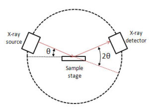
X-ray studies are mainly carried out in two basic configurations, namely, Single crystal and Powder XRD. However, the component parts of the x-ray spectrometer are in general common and comprise of:
- Source of x-rays
- Sample stage
- Detector
The article will provide basic details on the component parts of the x-ray diffractometer.
Source of x-rays

X-ray tube is a common source of x-rays. It comprises of an evacuated tube which contains a copper block anode bearing a metal target made of any of the metals such as molybdenum, tungsten, copper, rhodium, silver or cobalt .The cathode is a tungsten filament .On passage of electric current through the filament electrons are generated which move towards the anode under the highly accelerated voltage typically 30 – 150 kV. The accelerating electrons on striking the metal surface knock out electrons from the inner shells and the vacancies created are filled by electrons from the outer shells. In the process metal atoms emit x-rays. However, this involves heating of the metal block and x-rays constitute only a small fraction of the total energy liberated. The emitted x-rays exit the tube through a berylium window. The copper block needs to be cooled with a supply of water to dissipate the excessive heat generated. The Be window helps transmit a monochromatic beam of x-rays. Further monochromatization can be achieved by making use of a zirconium filter when using molybdenum as metal target. It absorbs the unwanted emissions while allowing the desired wavelengths to transmit.
Sample stage
Sample stage is also known as sample holder or a goniometer. Single crystal diffractometers make use of 4 circle goniometers. These circles help position the crystal planes for optimum x-ray diffraction settings. The sample stage can be a simple needle that holds the crystal in place or glass plate or fiber on which the crystal is mounted using an epoxy resin. Only sufficient quantity of epoxy resin is used so that the crystal is clearly mounted and not embedded in the resin. The fiber is mounted on a brass mounting pin and then inserted into the goniometer head. The sample is then centred with an optical arrangement such as a microscope or video camera and making adjustments along X, Y and Z directions to achieve optimum centering under the crosshairs of the viewer.
Detectors
In earlier days photographic films were used for recording the absorption pattern of diffracted beams. With the advances in detection technology more sensitive detector options were incorporated in advanced instruments. Such detectors include gas filled transducers, scintillation counters and semiconductor transducers. Solid state detectors offer highest levels of sensitivity and speed of analysis.
Subsequent articles will cover the operating principles and benefits of single crystal and powder systems.


